Medical & Surgical Nursing · Integumentary Function

Integumentary Function

Scabiesdefinition

Contagious ectoparasitic skin infestation caused by Sarcoptes scabiei.

in simple words: Spreads by prolonged skin-to-skin contact; outbreaks in crowded settings.

Scabies burrow – Pathognomonic sign

  • White, tortuous, thread-like lines
  • Between fingers, axillae, buttocks, elbows, ankles
  • Female mite in stratum corneum
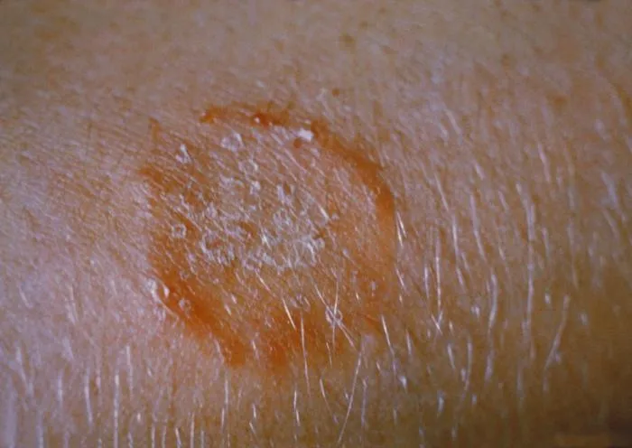
Circular scaly plaque with a raised red border and central clearing on the skin, typical of tinea corporis.
Tinea corporis ring lesion
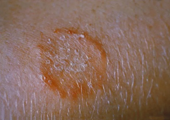
Well-demarcated annular erythematous scaly patch on the trunk consistent with tinea corporis.
Tinea corporis on skin

Scabies management

  • Treat all contacts – All household members and close contacts treated simultaneously, even if asymptomatic.
  • Wash bedding – Wash clothing, towels, bedding used in last 3 days in hot water ≥50°C and dry on high heat.
  • Burrow Ink Test – Ink applied to lesion and wiped away reveals serpentine burrow; quick bedside test.
© NextNurse

nextnurse.in

Integumentary Function · Medical & Surgical Nursing

Scabies & Tinea (contd.)

Tinea infections

TypeSiteFeatures
Tinea corporisBodyAnnular lesion with active erythematous scaly border and central clearing
Tinea crurisGroinJock itch; itching and redness in inguinal area
Tinea pedisFeetAthlete's foot; itching, scaling, fissures between toes
Tinea unguiumNailsThickened, brittle, discolored, crumbly nails

© NextNurse

nextnurse.in

Integumentary Function · Medical & Surgical Nursing

Wound Assessment

Wound infectiondefinition

Pus, redness, pain, swelling, hardness at wound site.

in simple words: Purulent drainage is definitive.

Granulation tissuedefinition

Pink or beefy red, moist, bumpy tissue indicating healthy healing.

in simple words: Bleeds easily, no pus. Not infection.

Infection

  • Pus, redness, pain, swelling
  • Purulent drainage

Granulation

  • Pink/beefy red, moist, bumpy
  • Bleeds easily, no pus

Pressure injury staging

StageDescription
1Intact skin, non-blanchable redness
2Partial-thickness loss; shallow ulcer or serum blister
3Full-thickness loss; fat visible, no bone/tendon/muscle
4Full-thickness loss; bone, muscle or tendon exposed
UnstageableSlough or eschar covers base
Deep tissue injuryNon-blanchable deep red, maroon or purple intact skin

© NextNurse

nextnurse.in

Integumentary Function · Medical & Surgical Nursing

Wound Assessment (contd.)

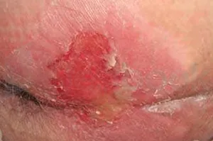
Pressure ulcer with partial-thickness skin loss presenting as a shallow open wound with a red-pink wound bed.
Stage 2 pressure ulcer
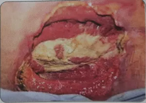
Deep pressure ulcer with full-thickness tissue loss exposing underlying structures such as muscle or bone.
Stage 4 pressure ulcer
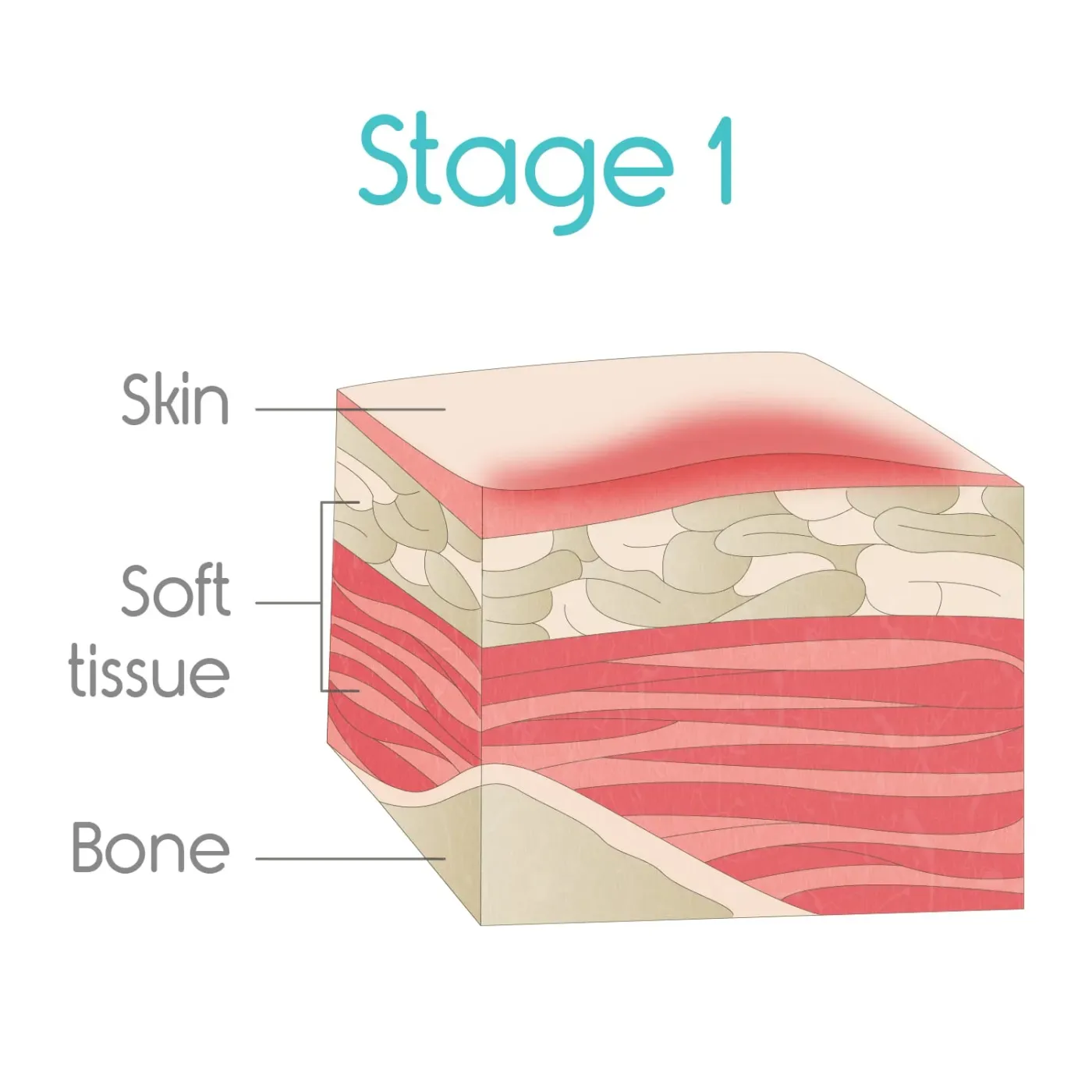
Intact skin over a bony prominence showing non-blanchable erythema, indicating a stage 1 pressure injury.
Stage 1 pressure ulcer

Why pressure injuries occur

Prolonged pressure over a bony prominence exceeds capillary closing pressure, obstructing blood flow and causing ischemia. Lack of oxygen and nutrients leads to cell death and ulcer formation.

© NextNurse

nextnurse.in

Integumentary Function · Medical & Surgical Nursing

Wound Healing Phases

Phases of wound healing

  1. Inflammatory1–4 days; haemostasis, clear bacteria/debris via neutrophils and macrophages
  2. ProliferativeDay 3–4 to weeks; fibroblasts synthesize collagen (Type III), granulation tissue forms
  3. MaturationReorganises collagen, replaces Type III with stronger Type I; increases tensile strength

Key points

Inflammatory phase does not synthesise collagen; macrophages release growth factors that stimulate later collagen production. Collagen synthesis occurs in proliferative phase. Maturation phase does not involve new collagen synthesis.

Phases

IPM

  • Inflammatory
  • Proliferative
  • Maturation

© NextNurse

nextnurse.in

Integumentary Function · Medical & Surgical Nursing

Pressure Injury Prevention

Repositioning

Reposition an immobile patient at least every 2 hours to relieve capillary compression. Risk assessment tools such as the Braden Scale guide frequency. Mobility is protective; immobility is a major risk factor.

Risk factors

  • Malnutrition – Impairs tissue repair; inadequate protein intake is a major cause of impaired healing.
  • Decreased sensory perception – Prevents pressure relief.
  • Incontinence – Moisture macerates skin.

Prevention measures

MeasureRationale
Reposition every 2 hoursRelieves capillary compression
Use pressure-redistribution surfaces (air mattresses)Distribute weight over larger area, lowering pressure intensity
Avoid hard bedsConcentrate pressure on bony prominences
Do not massage reddened prominencesCauses friction and shear, damages fragile capillaries
Keep skin clean and dryPrevents maceration from incontinence
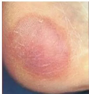
Non-blanchable erythema of intact skin over a bony prominence, the hallmark of a stage 1 pressure injury.
Stage 1 pressure injury

© NextNurse

nextnurse.in

Integumentary Function · Medical & Surgical Nursing

Wound Care & Dressings

Dressing selection

Dressing typeIndicationRationale
AbsorptiveModerate to heavy drainageRemoves exudate, prevents maceration
Hydrogel or transparent filmLittle to no drainageDonates moisture or protects wound bed
Sterile moist dressingBarrier against contaminationPrevents infection, supports epithelialization

Irrigation

Irrigate pressure ulcers with normal saline during each dressing change, not on a fixed schedule like every shift. Normal saline is isotonic and non-cytotoxic. Frequency depends on exudate and wound assessment. Irrigating every shift is too frequent and can traumatise granulation tissue and lower wound bed temperature.

Debridementdefinition

Removes dead, nonviable tissue from a wound so healing can proceed.

in simple words: May be mechanical, autolytic, chemical, or surgical. Necrotic tissue left in place prevents healing and predisposes to infection. Irrigation cleans but does not remove established necrotic tissue.

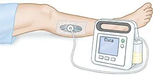
Negative pressure wound therapy

  1. SystemVacuum pump, sealed dressing, tubing, collection canister
  2. ActionDraws fluid, reduces swelling, stimulates new tissue growth
  3. AlarmCheck for air leak in dressing seal, kinked tubing, or full canister
Wound covered with a sealed foam dressing connected by tubing to a vacuum pump for negative pressure wound therapy.
Negative pressure wound dressing

© NextNurse

nextnurse.in

Integumentary Function · Medical & Surgical Nursing

Burns: Assessment

Rule of Nines (adults)

Body partPercentage
Head and neck9%
Each arm9%
Anterior trunk18%
Posterior trunk18%
Each leg18%
Perineum1%

Important

The Rule of Nines estimates TBSA, not burn depth or fluid volume. It is inaccurate in infants and young children because of their larger head and smaller legs; the Lund-Browder chart is preferred. The palm method estimates small burns: patient's palm ≈1% TBSA.

Burn depth classification

DegreeDepthFeatures
First-degreeEpidermis onlyErythema, pain like sunburn, no blisters
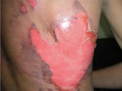
Second-degreeEpidermis and portion of dermisBlisters, redness, severe pain
Third-degreeFull-thickness, into subcutaneous tissueCharred, white, or leathery; dry, no blisters; painless
Fourth-degreeExtends into fascia, muscle, bonePainless; all skin layers including nerve endings destroyed
Skin burn with blistering and moist reddened surface indicating partial-thickness burn injury.
Partial thickness burn

© NextNurse

nextnurse.in

Integumentary Function · Medical & Surgical Nursing

Burns: Fluid Resuscitation

Parkland formuladefinition

4 mL × body weight in kg × %TBSA of lactated Ringer's in first 24 hours.

in simple words: Half given in first 8 hours from time of injury; remaining half over next 16 hours. The 8-hour clock starts at time of injury, not hospital admission.

Fluid choice

Hartmann's solution (lactated Ringer's) is the preferred isotonic crystalloid for initial fluid resuscitation in burns. Its electrolyte composition resembles plasma, and its lactate is metabolised to bicarbonate, correcting metabolic acidosis. 5% dextrose in water is not used because it becomes hypotonic and does not expand intravascular volume. Colloids are avoided in first 24 hours because increased capillary permeability lets proteins leak into interstitium and worsen edema.

Monitoring

ParameterTargetAction if below
Hourly urine output0.5–1 mL/kg/hrIncrease IV fluid rate as per protocol and notify doctor

© NextNurse

nextnurse.in

Integumentary Function · Medical & Surgical Nursing

Burns: Emergency Care

ABCDE protocol

  1. AirwayFirst priority, especially for facial, neck, or upper torso burns; signs of inhalation injury warrant early intubation
  2. BreathingAssess for inhalation injury; oxygen therapy
  3. CirculationFluid resuscitation to maintain circulation; Ringer's lactate
  4. DisabilityNeurological assessment
  5. ExposureRemove jewelry and constricting clothing

Chemical burns

  • Immediate irrigation – Copious water irrigation for at least 20–30 minutes to dilute and remove chemical.
  • Dry chemicals – Brush off powder before irrigating with water to prevent exothermic reaction.
  • Do not neutralize – Neutralization can generate heat and worsen injury.

© NextNurse

nextnurse.in

Integumentary Function · Medical & Surgical Nursing

Burns: Complications

Circumferential burns

A circumferential burn can cause airway obstruction, poor respiratory effort, or absent peripheral pulses depending on its location. Neck or face burns cause airway obstruction from swelling; chest burns restrict chest wall movement; limb burns compress vessels and cause compartment syndrome. Emergency escharotomy is a surgical incision through the eschar to relieve pressure and restore breathing or circulation.

Neurovascular checks: 6 Ps

  • Pain – Assess for pain
  • Pallor – Check color
  • Pulselessness – Palpate pulses
  • Paresthesia – Check sensation
  • Paralysis – Assess movement
  • Poikilothermia – Check temperature

Electrolyte imbalances

ImbalanceCauseTiming
HyperkalemiaRelease of potassium from damaged cellsEarly emergent phase
HypokalemiaFluid shifts back into vascular space and diuresis48–72 hours post-burn
Metabolic acidosisDecreased tissue perfusion and lactic acid productionEarly phase

© NextNurse

nextnurse.in

Integumentary Function · Medical & Surgical Nursing

Burns: Special Considerations

Inhalation injurydefinition

Determines the need for ventilatory assistance, not burn surface area.

in simple words: Signs include facial burns, singed nasal hairs, soot in the airway, and hoarseness. Early intubation may be required.

Electrical burns

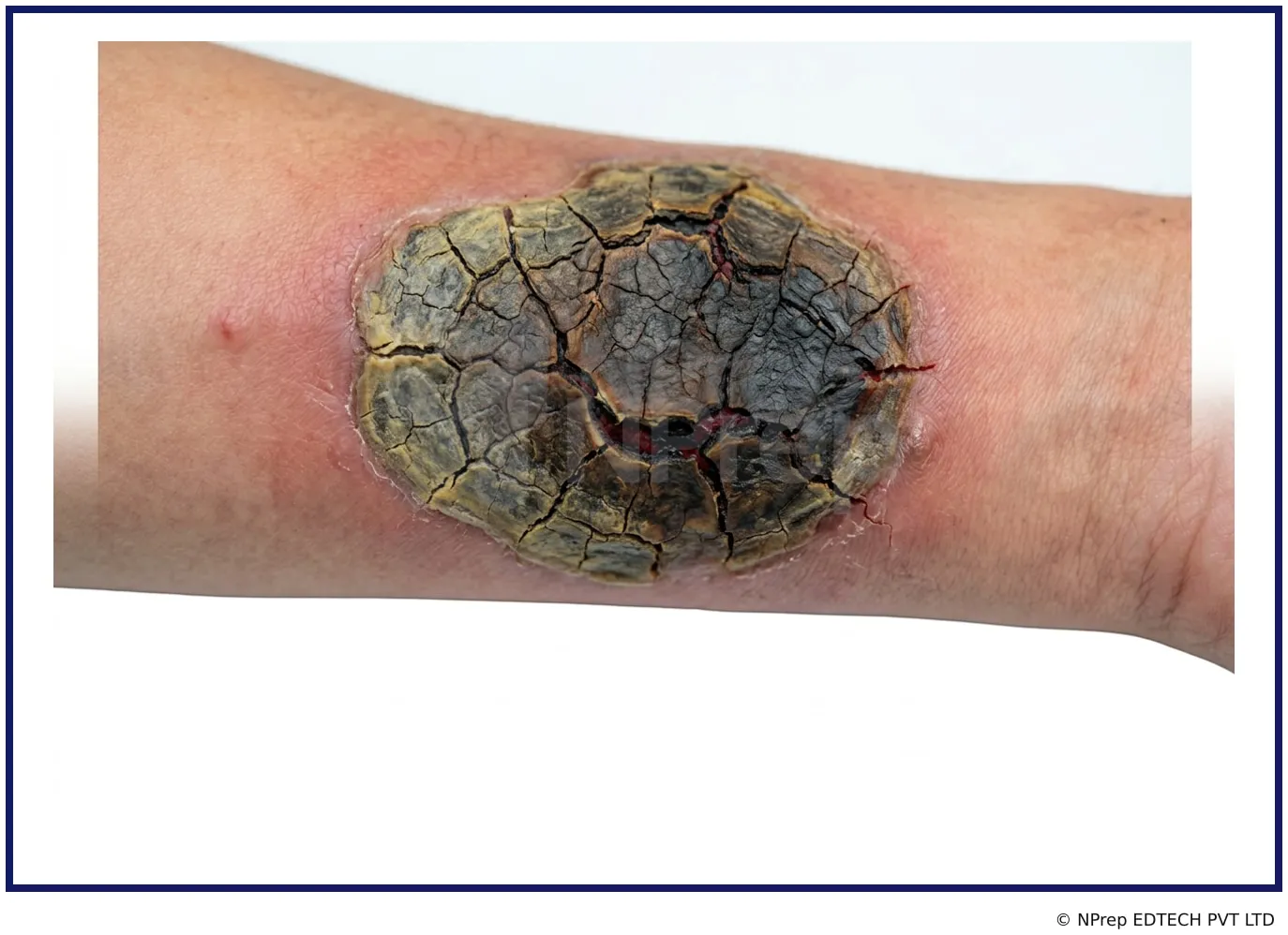
High-voltage electrical burns produce a hard, dry, leathery eschar at entry and exit points, described as crocodile-like skin. The current generates extreme heat, causing coagulation necrosis and full-thickness injury that destroys nerve endings, making the area painless.

Hard dry leathery charred skin with crocodile-like cracking seen in high-voltage electrical burns.
Crocodile skin electrical burn

Facial burns

Facial burns are classified as major injuries requiring immediate hospital admission regardless of TBSA. The primary risk is airway compromise from edema of the face, neck, and upper airway, which can develop hours after injury even in a conscious patient. Signs of developing airway obstruction include hoarseness, stridor, sooty sputum, and difficulty swallowing.

© NextNurse

nextnurse.in

Integumentary Function · Medical & Surgical Nursing

Skin Infections & Disorders

Cellulitisdefinition

Diffuse, spreading bacterial infection of the dermis and subcutaneous tissue.

in simple words: Common organisms: Streptococcus pyogenes and Staphylococcus aureus. Presents with erythema, oedema, warmth, tenderness, poorly demarcated borders.

Impetigodefinition

Superficial skin infection of the epidermis, often caused by Staphylococcus aureus.

in simple words: Presents as honey-coloured crusts; more superficial than cellulitis.

Paronychiadefinition

Localised infection of the skin fold around a fingernail or toenail.

in simple words: A felon is a deep abscess of the fingertip pulp. Both are localised, unlike cellulitis which spreads diffusely.

Carbuncledefinition

Deep infection of multiple interconnected boils (furuncles) arising from hair follicles.

in simple words: Localised abscess rather than diffuse soft tissue infection seen in cellulitis.

© NextNurse

nextnurse.in

Integumentary Function · Medical & Surgical Nursing

Autoimmune & Other Disorders

Pemphigusdefinition

Autoimmune disorder causing acantholysis and flaccid, painful bullae that rupture easily.

in simple words: Autoantibodies attack desmogleins, leading to loss of cell adhesion; oral lesions common and painful. Positive Nikolsky sign.

Bullous pemphigoiddefinition

Presents with tense blisters, intense itching, and a negative Nikolsky sign.

in simple words: Unlike pemphigus, blisters are subepidermal and do not rupture easily; anti-pruritic treatment is a priority.

Plaque psoriasisdefinition

Well-demarcated erythematous plaques with shiny, silvery-white adherent scales.

in simple words: Typically on extensor surfaces such as elbows, knees, scalp, lower back. Driven by autoimmune hyperproliferation of keratinocytes.

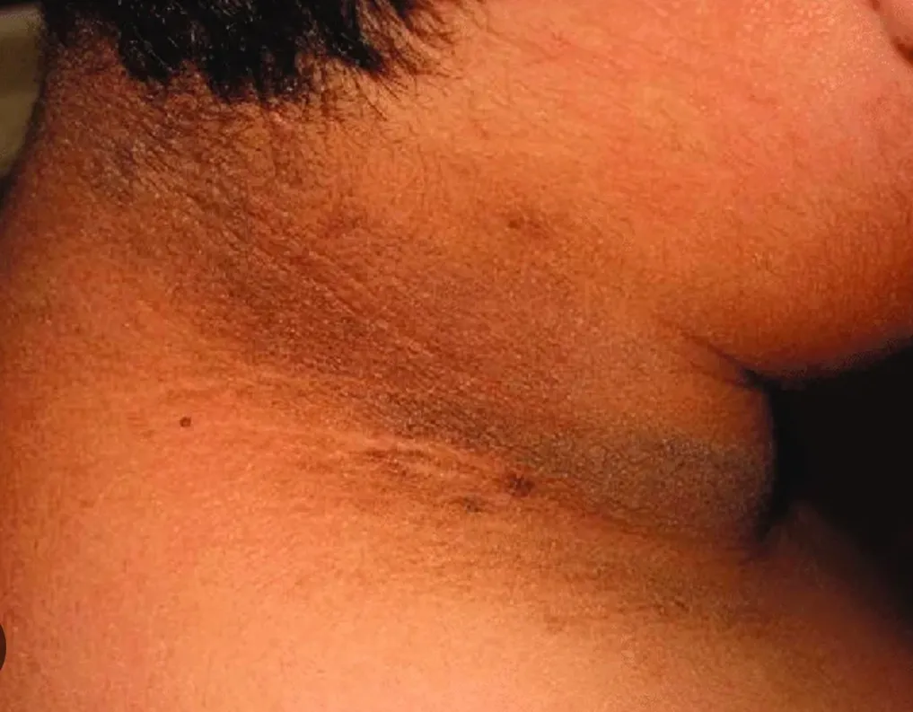
Acanthosis nigricansdefinition

Velvety, hyperpigmented, thickened plaques in intertriginous areas, especially posterior neck and axillae.

in simple words: Strongly associated with insulin resistance and diabetes mellitus; clinical marker for metabolic syndrome.

Velvety hyperpigmented thickened plaques on the posterior neck and flexural folds typical of acanthosis nigricans.
Acanthosis nigricans

© NextNurse · Checked against: NextNurse Q-Bank explanations

nextnurse.in