Nursing Foundations · Diagnostic testing

Diagnostic Testing

Diagnostic specimen collectiondefinition

Obtaining a representative sample of body fluid or tissue for laboratory analysis, using aseptic technique and correct timing to ensure accurate results.

in simple words: Collect the right sample, at the right time, in the right way.

Sputum for culture – Best time: early morning, immediately after waking

  • Collect before oral hygiene, food or drink.
  • Plain water rinsing is allowed.
  • Must be coughed up from lungs, not saliva.
  • If unable: steam inhalation, aerosol therapy, chest physiotherapy may help mobilise.
  • High yield

Midstream clean-catch urine – Standard for urine culture & sensitivity

  • Discard first stream (flushes urethral flora).
  • Collect midstream in sterile container.
  • Minimum 10 mL required.
  • Do not collect from drainage bag.
  • High yield

© NextNurse

nextnurse.in

Diagnostic Testing · Nursing Foundations

Blood Collection Order

Why order matters

The sequence of draw prevents additive carryover between tubes. Blood culture tubes are drawn first to maintain sterility; citrate tubes next because other additives alter clotting times; heparin tubes follow; oxalate tubes last because their additives interfere with electrolyte and coagulation tests.

Correct order of draw

  1. 1. CultureSterile; drawn first to prevent contamination.
  2. 2. Citrate (light blue)For PT, aPTT, fibrinogen. Underfilling falsely prolongs PT/INR.
  3. 3. Heparin (light green)For plasma chemistry, not chromosome/HLA testing.
  4. 4. Oxalate (grey)For glucose & lactate. Drawn last to avoid interference.

Tube additives & uses

Tube topAdditiveUse
Light blueSodium citrateCoagulation studies (PT, aPTT, fibrinogen)
GreySodium fluoride + potassium oxalateGlucose, lactate
Light greenHeparinPlasma chemistry
RedNoneSerum chemistry; glycolysis continues if used for glucose

© NextNurse

nextnurse.in

Diagnostic Testing · Nursing Foundations

Urine & Stool Tests

24-hour urine collection – Quantitative analysis

  • Used for hormones, proteins, creatinine clearance.
  • Not for bacterial culture (not sterile).
  • Discard first void; start timing then.
  • If a void is missed, test is invalid — restart.

First-morning midstream urine – Routine urinalysis & microscopy

  • Concentrated overnight — increases detection of cells, casts, bacteria, protein.
  • Midstream avoids urethral contamination.
  • Clean genital area with antiseptic towelette, not alcohol swab.

Stool & other tests

  • Guaiac FOBT – Detects hidden blood in stool; heme acts as pseudoperoxidase, turns guaiac blue.
  • Fouchet's test – Detects bile pigments in urine, not blood in stool.
  • Sweat chloride – Gold standard for cystic fibrosis; ≥60 mmol/L confirms CF.
  • Fecal fat – Confirms steatorrhea but not specific to CF.

© NextNurse

nextnurse.in

Diagnostic Testing · Nursing Foundations

Visual & Ear Assessment

Tools for eye assessment

An ophthalmoscope views internal eye structures (retina, optic nerve) but does not measure visual acuity. Visual acuity is tested with the Snellen chart (distance) and Jaeger chart (near). Colour vision is screened with the Ishihara chart. Record acuity for each eye separately (OD, OS) and note corrective lenses.

Eye assessment tools

ToolPurpose
OphthalmoscopeView retina, optic nerve
Snellen chartDistance visual acuity (20 feet / 6 m)
Jaeger chartNear vision
Ishihara chartColour vision deficiency

Otoscopy: Adult – Straighten ear canal

  • Pull pinna upward and backward.
  • Visualises tympanic membrane (pearly grey, translucent).

Otoscopy: Infant/child <3 yr – Straighten ear canal

  • Pull pinna downward and backward.
  • Adult technique would curve canal further, risking injury.
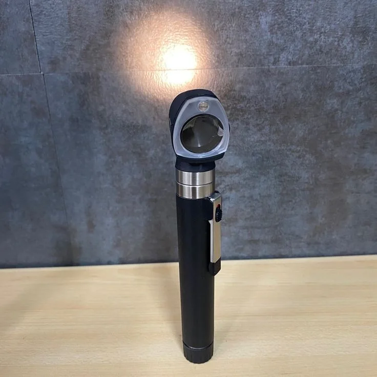
A healthcare provider using an otoscope to examine a patient's ear.
Otoscope for ear examination

Image: Fatimah Bello, CC0

© NextNurse · Checked against: NextNurse Q-Bank explanations

nextnurse.in